Facilitating insights into life
Users of imaging systems in Life Science Research have a broad range of different imaging needs enabling them to address a wide variety of different applications. Cameras support users in all life science imaging workflows: from cell culture checking, documentation of stained specimens for morphological examinations to live cell imaging and analytical methods such as FRAP and FRET. They generate reliable, reproducible, quantifiable data that can be used for analyses and comparison.
Tools for all researchers
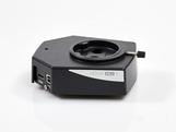
Leica microscope cameras are scientific tools that provide precise monitoring and documentation of specimens and tissues. They support all transmitted light contrasting methods including Differential Interference Contrast (DIC), Phase Contrast (PH), and Brightfield (BF). To achieve publication-ready results, their color reproduction perfectly matches all kinds of staining procedures.
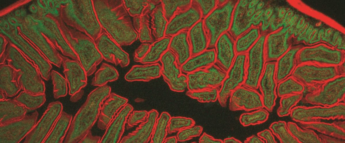
For fluorescence applications, cameras should capture as many photons emitted from the fluorescent specimen as possible whilst introducing little additional noise to the data. Leica Microsystems offers a range of color or monochrome fluorescence cameras based on CCD or sCMOS sensors. They are designed to address basic applications such as documentation of fixed immuno-stained specimens through to demanding real-time, high-speed triggered, multidimensional acquisitions.


