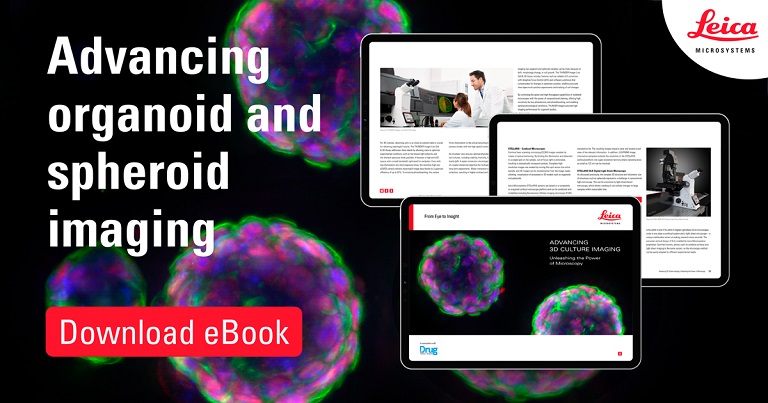
Advancing 3D culture imaging, Unleashing the power of microscopy!
This eBook examines some of the challenges encountered when imaging 3D culture models and presents case studies where innovative microscopy solutions have made new research methods possible.
Case studies presented:
- Studying human brain development and disease examining “brains-in-a-dish” from induced pluripotent stem cells (iPSCs)
- Observing 3D cell cultures during development
- Developing heart pacemaker cells from cardiac spheroids
- Imaging anti-cancer drug uptake in spheroids using DLS microscopy
- Faster 3D imaging of cellular and protein structures

Curious about the image?
Murine oesophageal organoids (DAPI, Integrin26-AF 488, SOX2-AF568) imaged with a THUNDER Imager 3D Cell Culture. Courtesy of Dr. F.T. Arroso Martins, Tampere University, Finland.
